Home
Uncategories
Back Muscle Diagrams Labeled - Deep Back Muscles Anatomy Innervation And Functions Kenhub / Does degenerative disc disease affect the lower back muscle?
Back Muscle Diagrams Labeled - Deep Back Muscles Anatomy Innervation And Functions Kenhub / Does degenerative disc disease affect the lower back muscle?
Back Muscle Diagrams Labeled - Deep Back Muscles Anatomy Innervation And Functions Kenhub / Does degenerative disc disease affect the lower back muscle?. This article looks at the anatomy of the back, including bones, muscles, and nerves. Does degenerative disc disease affect the lower back muscle? Labeled vector back muscles labeled anatomical educational body scheme vector illustration. Black and white muscular system diagram label muscles. Start studying 08 back muscles label.
They start at the top of the neck and go down to the tailbone. Superficial back muscles, intermediate back muscles and intrinsic back muscles.the intrinsic muscles are named as such because their embryological development begins in the back, oppose to the superficial and intermediate back muscles which develop elsewhere and are therefore classed as extrinsic muscles. Whatever you need, whatever you want, whatever you desire, we provide. Back pain is one of the most common kinds of pain for adults, and muscle strains are the most common type of back pain. For more anatomy content please follow us and visit our website:
Muscle Anatomy Hd Stock Images Shutterstock from image.shutterstock.com Vertebrae, bones, joints, ligaments, muscles, muscular system, fascia, arteries, veins, nerves and various adjacent organs. Does degenerative disc disease affect the lower back muscle? Muscle or ligament strains can occur from repeated use of the muscles, or from improperly or awkwardly lifting heavy objects. The back anatomy includes the latissimus dorsi, trapezius, erector spinae, rhomboid, and the teres major. Search for muscle anatomy diagram. Broadly considered, human muscle—like the muscles of all vertebrates—is often divided into striated muscle, smooth muscle, and cardiac muscle. Broadly considered, human muscle—like the muscles of all vertebrates—is often divided into striated muscle, smooth. The back anatomy includes the latissimus dorsi, trapezius, erector spinae, rhomboid, and the teres major.
Back pain is common and might be caused by a problem with a muscle.
Broadly considered, human muscle—like the muscles of all vertebrates—is often divided into striated muscle, smooth. It also covers some common conditions and injuries that can affect the back. On this page, you'll learn about each of these muscles, their locations and functional anatomy. Muscles of lower back diagram in this image, you will find an occipital bone, sternocleidomastoid, trapezius, deltoid in muscles of the lower back diagram. The muscles of the back. The external intercostal muscles, or external intercostals (intercostales externi) are eleven in number on both sides. Black and white muscular system diagram label muscles. Shoulder muscle anatomy human muscle anatomy human anatomy chart human anatomy and physiology lower back anatomy muscle diagram anatomy organs anatomy images scoliosis exercises. It comprises the vertebral column (spine) and two compartments of back muscles; Lower back muscle diagram anatomy. Anatomynote.com found anatomy of back muscles diagram from plenty of anatomical pictures on the internet. Many in the neck help to stabilize or move the head. Back pain is common and might be caused by a problem with a muscle.
Anatomynote.com found anatomy of back muscles diagram from plenty of anatomical pictures on the internet. By the way, have you heard about the myth of. They start at the top of the neck and go down to the tailbone. Extrinsic and intrinsic.the back functions are many, such as to house and protect the spinal cord, hold the body and head upright, and adjust the movements of the upper and lower limbs. Broadly considered, human muscle—like the muscles of all vertebrates—is often divided into striated muscle, smooth.
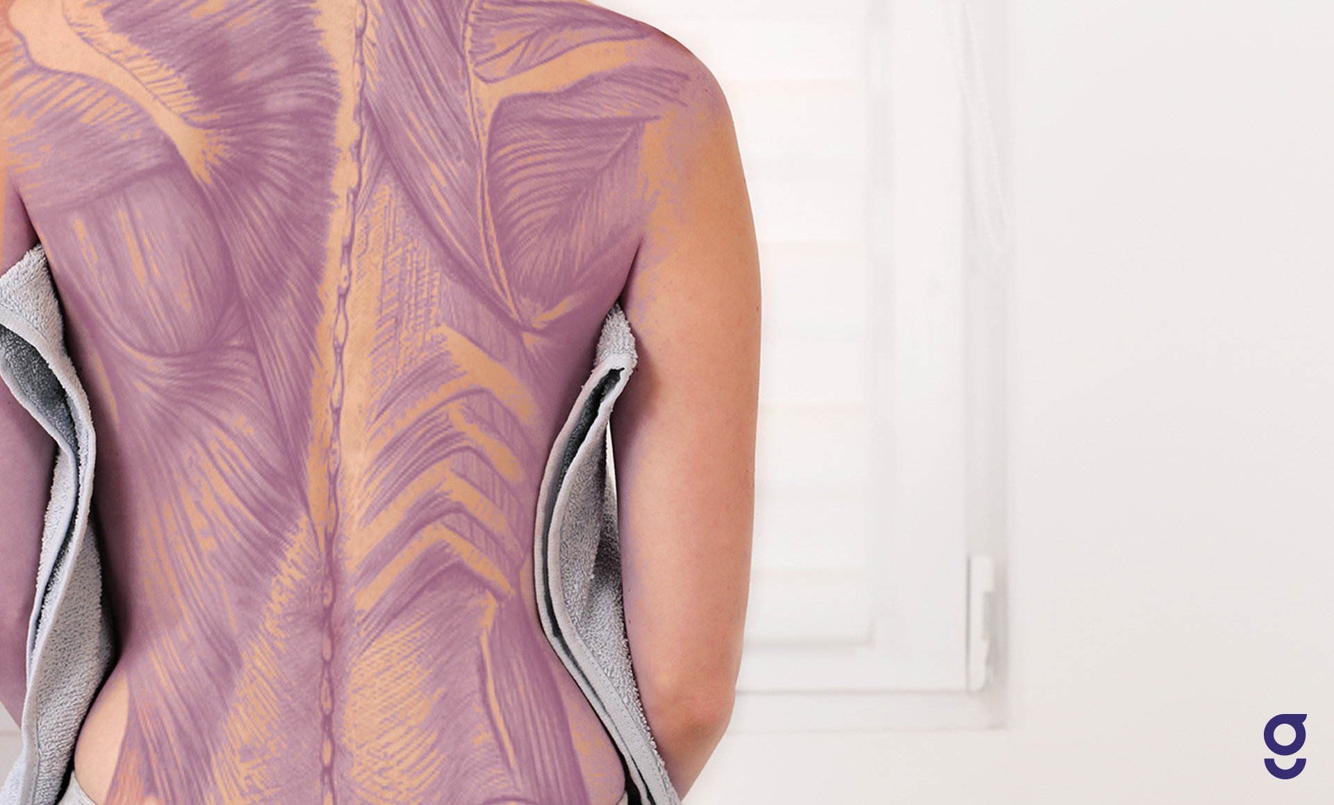
Back Muscles Anatomy Of Back Pain In Diagrams Goodpath from images.ctfassets.net Start studying 08 back muscles label. Three types of back muscles that help the spine function are extensors, flexors and obliques. Does degenerative disc disease affect the lower back muscle? Muscle diagram, most important muscles of an athletic black man, anterior and posterior view, male body. The back is the body region between the neck and the gluteal regions. On this page, you'll learn about each of these muscles, their locations and functional anatomy. To learn more about the anatomy of the spine, watch this video. The muscles of the back can be arranged into 3 categories based on their location:
The back anatomy includes the latissimus dorsi, trapezius, erector spinae, rhomboid, and the teres major.
On this page, you'll learn about each of these muscles, their locations and functional anatomy. It comprises the vertebral column (spine) and two compartments of back muscles; We have a collection of back muscle diagrams to help you learn more about the structure of muscles on your back. Back muscle diagram back muscles big back big back muscles big lats bodybuilding secrets major back muscles. Nerves in your lower back. Muscles make up a large part of the anatomy (structure) of the back. We are pleased to provide you with the picture named anatomy of back muscles diagram.we hope this picture anatomy of back muscles diagram can help you study and research. On anatomical parts the user can choose to display the various structures in colored illustrations of the anatomy of the back and spine: Related posts of back muscle diagrams labeled muscle anatomy in thigh. As you can see, there are also have a spine of scapula deltoid, triceps brachii, latissimus dorsi. To learn more about the anatomy of the spine, watch this video. This is a diagram of the larger and more surface muscles of the low back. Back pain is one of the most common kinds of pain for adults, and muscle strains are the most common type of back pain.
Using this atlas of human anatomy of the spine and back. This picture also contains humerus, olecranon process of ulna, deep to tendon and so on. Whatever you need, whatever you want, whatever you desire, we provide. Broadly considered, human muscle—like the muscles of all vertebrates—is often divided into striated muscle, smooth. Facebook twitter google+ linkedin stumbleupon tumblr pinterest reddit vkontakte share via email print.
Free Anatomy Quiz The Muscular System Section from www.free-anatomy-quiz.com To learn more about the anatomy of the spine, watch this video. Labeled sti infection development diagram. It also covers some common conditions and injuries that can affect the back. Muscle anatomy in thigh 12 photos of the muscle anatomy in thigh muscle anatomy inner thigh, muscle anatomy of thigh, muscle anatomy of upper thigh, muscle anatomy posterior thigh, muscle anatomy thigh mri, human muscles, muscle anatomy inner thigh, muscle anatomy of thigh, muscle anatomy of upper thigh, muscle anatomy. Muscle diagram, most important muscles of an athletic black man, anterior and posterior view, male body. Muscles labeled front and back / probes / muscles vary greatly in their shape and size. Back muscle diagram back muscles big back big back muscles big lats bodybuilding secrets major back muscles. Related posts of back muscle diagrams labeled muscle anatomy in thigh.
Back pain is common and might be caused by a problem with a muscle.
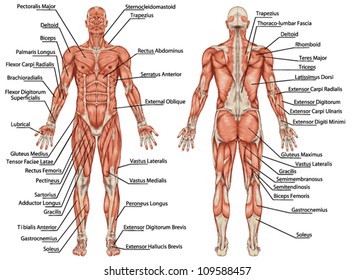
Muscle anatomy in thigh 12 photos of the muscle anatomy in thigh muscle anatomy inner thigh, muscle anatomy of thigh, muscle anatomy of upper thigh, muscle anatomy posterior thigh, muscle anatomy thigh mri, human muscles, muscle anatomy inner thigh, muscle anatomy of thigh, muscle anatomy of upper thigh, muscle anatomy. Anatomynote.com found anatomy of back muscles diagram from plenty of anatomical pictures on the internet. Using this atlas of human anatomy of the spine and back. Another common cause of lower back and hip pain is disc injury. The external intercostal muscles, or external intercostals (intercostales externi) are eleven in number on both sides. Abdomen muscles labeled labeled diagram of muscles gallery: Muscles of lower back diagram in this image, you will find an occipital bone, sternocleidomastoid, trapezius, deltoid in muscles of the lower back diagram. Extrinsic and intrinsic.the back functions are many, such as to house and protect the spinal cord, hold the body and head upright, and adjust the movements of the upper and lower limbs. Search for muscle anatomy diagram. Black and white muscular system diagram label muscles. It also covers some common conditions and injuries that can affect the back. Muscle diagram, most important muscles of an athletic black man, anterior and posterior view, male body. This labeled human muscular system chart illustrates the major muscle groups in the back (posterior) view and the front (anterior) view.
On anatomical parts the user can choose to display the various structures in colored illustrations of the anatomy of the back and spine: back muscle diagram. Five pairs of lumbar spinal nerves labeled l1 to l5 branch off your spinal cord and exit through small holes between the vertebrae.

0 Comments:
Posting Komentar